Which Leads Reflect Ischemic Changes in the Right Coronary Artery
The culprit artery for this patient is the right coronary artery. Alternatively acute occlusion of the right coronary artery may produce ischemia in the anterior left ventricular wall supplied by a stenotic anterior descending coronary artery.

Characteristics And Outcomes Of Coronary Artery Involvement In Polyarteritis Nodosa Canadian Journal Of Cardiology
In group 2 the electrocardiographic changes reflect extensive subendocardial myocardial ischemia.

. Background Chest pain frequently occurs without any signs of ischemia within the first 24 hours after coronary interventions. Changes usually are seen in the leads that reflect the distribution of the diseased coronary artery. LBBB did not mask the ECG recognition of the occluded coronary artery during the first 60 min of ischemia but 3 hours later only the LAD occlusion could be reliably identified.
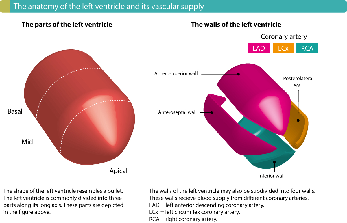
Left Coronary Artery Right Coronary Artery. Walls of the LV SA node in 55 of people AV node in 90 of people Posterior fascicle of the LBB Left Anterior Descending Artery LAD. Leads II III and aVF.
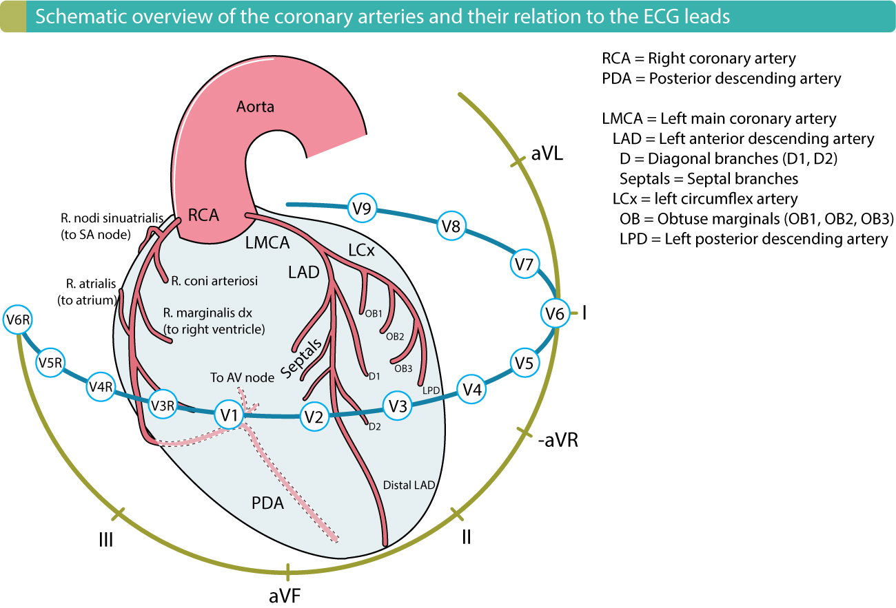
Occlusion of the right coronary artery RCA may cause infarction of the inferior wall of the left ventricle with or without right ventricular RV myocardial infarction MI manifested as ST-segment elevations in. Therefore in most people the changes seen in leads II III and AVF inferior leads reflect disease in the right coronary. However 10 of the time this arises in a branch of the left coronary artery and the remaining 20 display a dual origin.
The clot might block an artery and lead to sudden severe myocardial ischemia resulting in a heart attack. II III and aVF. Annual mortality rate from all causes across Europe amounts to 11 per 1000 inhabitants with 54 per 1000 49 due to cardiovascular disease and 24 per 1000 22 due to ischemic heart disease In patients suffering from coronary artery disease CAD the size of the myocardial infarction is the most important determinant of the outcome after such an event.
Left main and left circumflex coronary arteries were normal and the previously placed midleft anterior descending. Which leads reflect ischemic changes in the right coronary artery. An inferior wall MI also known as IWMI or inferior MI or inferior ST segment elevation MI or inferior STEMI occurs when inferior myocardial tissue supplied by the right coronary artery.
To test the hypothesis that this pain may be due to local vessel injury stretch pain we performed a prospective study enrolling patients after PTCA stent implantation or diagnostic coronary angiography alone. Early change suggestiveof AMI. This temporary tightening of the muscles in the artery wall can briefly decrease or even prevent blood flow to part of the heart.
Other features of RV hypertrophy may suggest this diagnosis eg terminal S-wave in lead I TWI in lead III. STEMI is defined electrocardiographically as an acute ST-segment elevation at the J-point in two contiguous leads with varying cut-points for sex and lead type in the setting of a clinical syndrome suggestive of myocardial ischemia. Three hours after coronary occlusion ST segment changes declined progressively and only the LAD occlusion could be reliably recognized.
The ECG changes illustrated here indicate ischemia during anginal pain. In a previous experimental study in pigs we found that simultaneous occlusion of the left anterior descending LAD and right coronary arteries attenuated the ST-segment elevation and blunted the reciprocal ST-segment depression in the 12-lead ECG Cinca et al 2014 but combination of double occlusions involving the three main coronary systems were not explored. A patient with electrocardiogram changes in leads V3 and V4 that reflect an infarction is experiencing an injury to which area of the heart.
Ischemia Injury Infarction. The increased frequency of ischemic changes noted on screening ECGs in patients with diabetes simply may reflect their greater baseline risk. The changes in the ST-segment reflect currents of injury elicited by potential gradients between ischemic and non-ischemic myocardium and the.
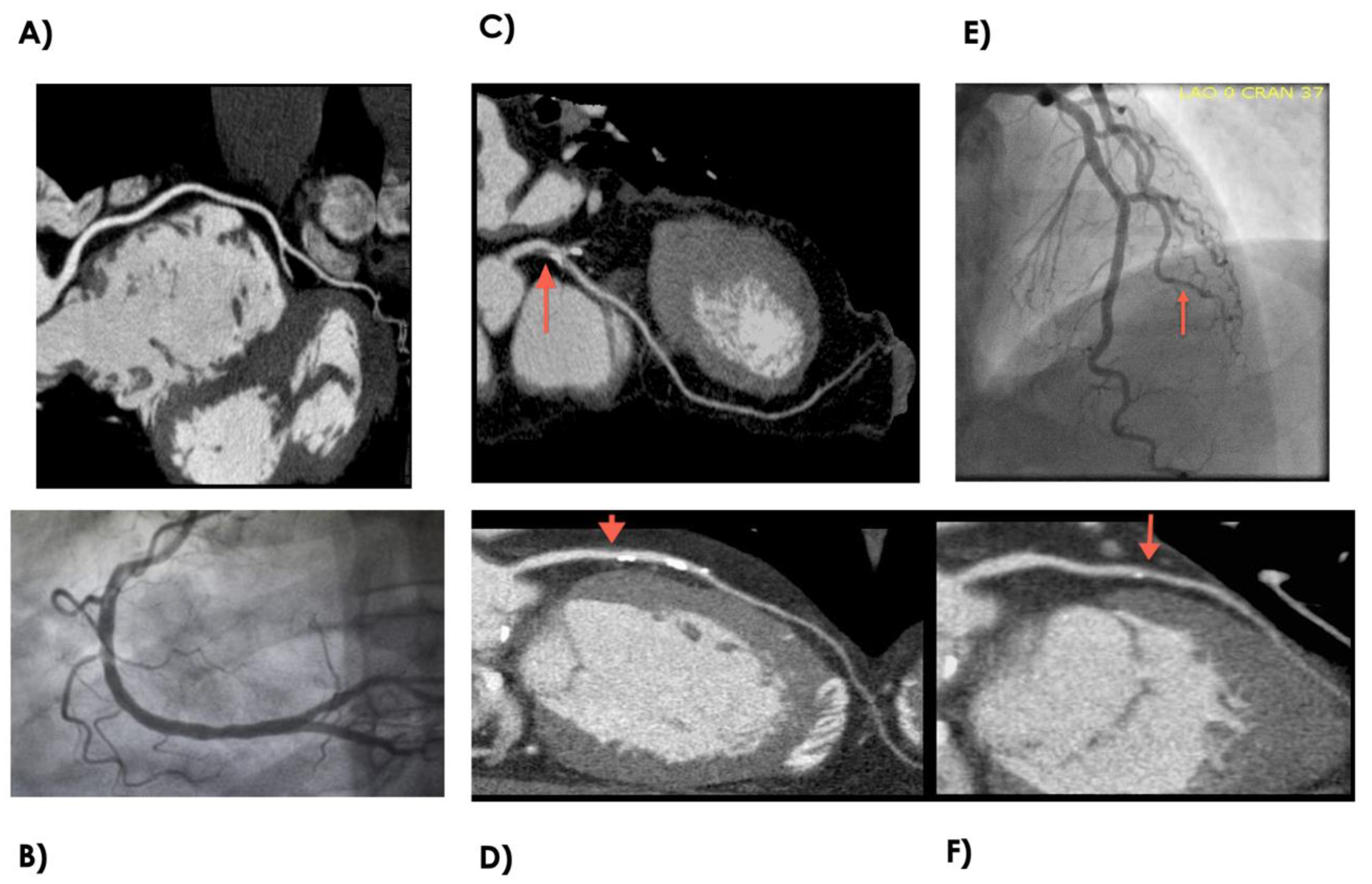
This ECG shows ST-segment elevation in the inferior wall leads. Vessel caliber increased from less than 2 mm to greater than 4 mm. Clues to suggest digoxin or hypokalemia.
In 70 of cases this artery indeed arises from the right coronary artery. Lead aVL or both Right coronary artery 90 71 94 70 Absence of the above findings plus ST-segment elevation in leads I aVL V 5 and V 6 and ST-segment depression in leads V 1 V 2 and V 3 Left. The right coronary artery is one of several major vessels that provide blood to the heart.
ST-segment elevations were noted in the right-sided leads along with a dramatic increase in ST-segment elevations in the traditional limb leads and reflected her clinical deterioration. 1 RV hypertrophy may cause a tall R-wave in the right precordial leads and a strain pattern in V1-V3 with STD. Rarely a blood clot might travel to the coronary artery from elsewhere in the body.
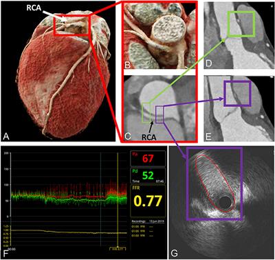
Coronary angiography showed a dominant right coronary artery RCA with 60 mid-vessel narrowing which decreased to less than 30 after administration of intracoronary nitroglycerin. Furthermore the posterior descending artery may be much smaller meaning that other structures. McHenry Western Lake County EMS.
Up to 7 cash back Page 5 See page 2 for important information about the uses and limitations of this guide and page 25 for all third-party sources. Ischemia and ST changes Coronary Arteries Mechanisms of ischemia Treatment Ischemia and MI EKG changes Right Coronary Artery RCA Supplies RA and RV Inf and post. Acute ST-segment elevations in the precordial leads generally suggest acute occlusion in the branches of the left coronary artery.
We found severe coronary artery disease LMCA or right main artery equivalent in 69 of such patients and the mortality in those with subsequent myocardial infarction was 77 mainly due to pump failure. ICD-10-CM1 Descriptor I25111 Atherosclerotic heart disease of native coronary artery with angina pectoris with documented spasm I25118 Atherosclerotic heart disease of native coronary artery with other forms of. Anterior ischemia may result from the abnormal hemodynamics or the reduced collateral flow produced by acute right coronary artery occlusion.
Which drug is used to manage a patient with a dissecting aneurysm. 2 Digoxin or hypokalemia may cause scooped STD in the right precordial leads. The right coronary artery splits into the acute marginal arteries and the right posterior coronary artery.
The right-sided ECG was performed after an initial attempt at stabilization in the cardiac intensive care unit and is shown in Figure 1. Origin of posterior descending artery. Patients with major stenoses of the left coronary system many of whom had suffered old inferior wall infarction showed ST changes in Leads I aVL and the chest leads.
Patients with major stenoses of the right coronary artery with or without disease of the left coronary system showed ischemic ST changes in Leads II III and aVF.

Coronary Heart Disease Health Information Bupa Uk
Invasive Management Of Coronary Artery Disease In Advanced Renal Disease Kidney International Reports

Arrhythmogenic Cardiomyopathy Dilated Cardiomyopathy Is A Condition In Which The Heart Becomes Cardiac Nursing Cardiovascular Nursing Medical
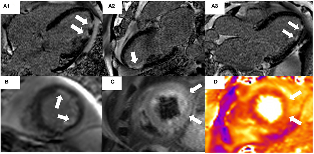
Frontiers The Role Of Cardiac Magnetic Resonance In Myocardial Infarction And Non Obstructive Coronary Arteries Cardiovascular Medicine
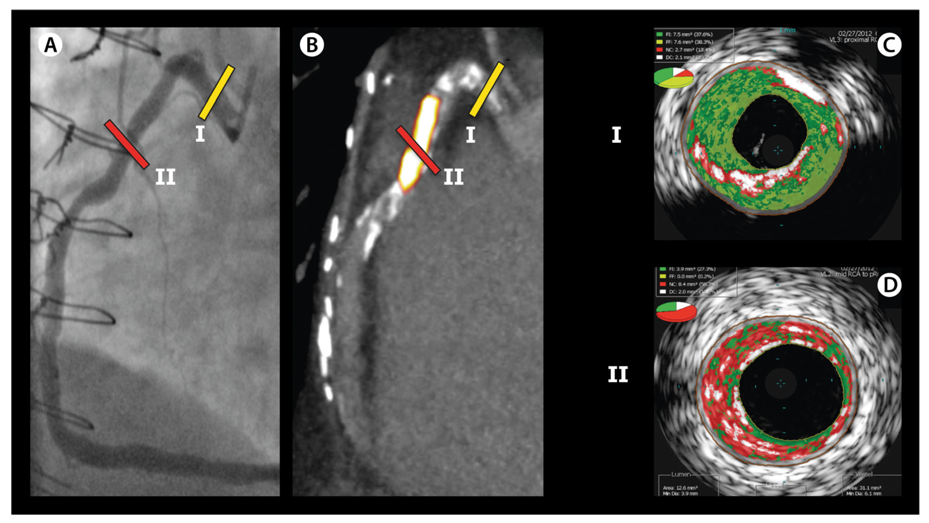
Jcm Free Full Text Detection Of Vulnerable Coronary Plaques Using Invasive And Non Invasive Imaging Modalities Html

Ecg Localization Of Myocardial Infarction Ischemia And Coronary Artery Occlusion Culprit Ecg Echo

Ecg Localization Of Myocardial Infarction Ischemia And Coronary Artery Occlusion Culprit Ecg Echo
2
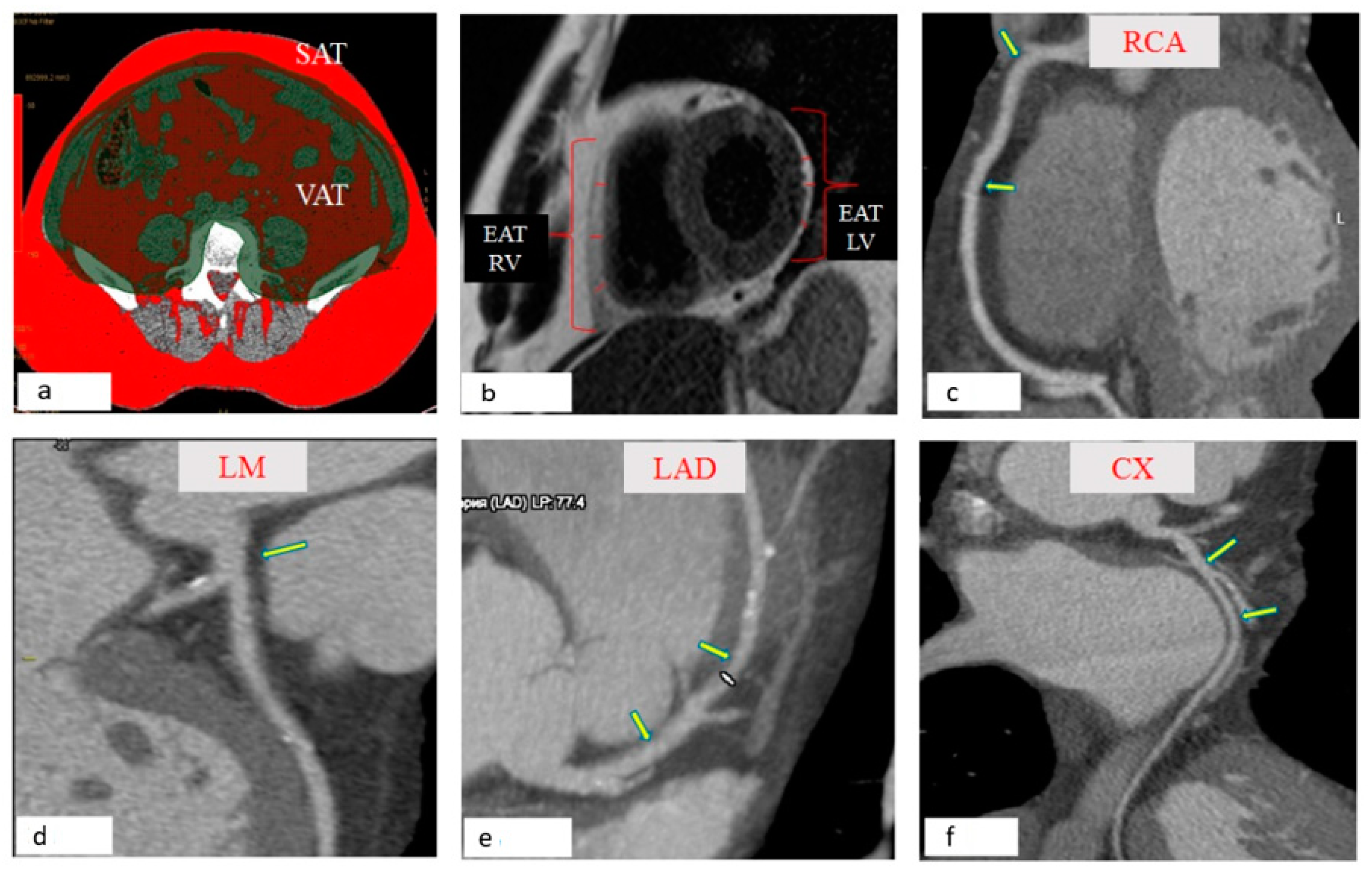
Jpm Free Full Text Relationship Between Epicardial And Coronary Adipose Tissue And The Expression Of Adiponectin Leptin And Interleukin 6 In Patients With Coronary Artery Disease Html

Pin On Ecg
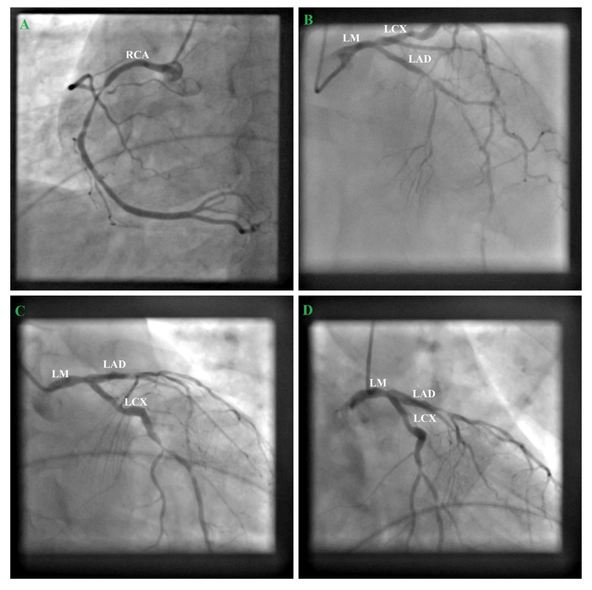
Medicina Free Full Text Severe Coronary Artery Disease In A Person Living With Hiv Html

Combined Assessment Of Subtended Myocardial Volume And Myocardial Blood Flow For Diagnosis Of Obstructive Coronary Artery Disease Using Cardiac Computed Tomography A Feasibility Study Journal Of Cardiology

Ecg The Acute Coronary Syndromes Ppt Video Online Download
Ecg Localization Of Myocardial Infarction Ischemia And Coronary Artery Occlusion Culprit Ecg Echo
2
Frontiers Hemodynamic Relevance Of Anomalous Coronary Arteries Originating From The Opposite Sinus Of Valsalva In Search Of The Evidence Cardiovascular Medicine
Diagnostics Free Full Text Early Detection Of Coronary Artery Disease By Micro Rna Analysis In Asymptomatic Patients Stratified By Coronary Ct Angiography Html
Ecg Localization Of Myocardial Infarction Ischemia And Coronary Artery Occlusion Culprit Ecg Echo

Surgical Anatomy 1 Ascending Aorta Cannulation Sites On The Ascending Aorta Should Be As High Abdominal Aorta Cardiovascular System Vein Thrombosis


Comments
Post a Comment